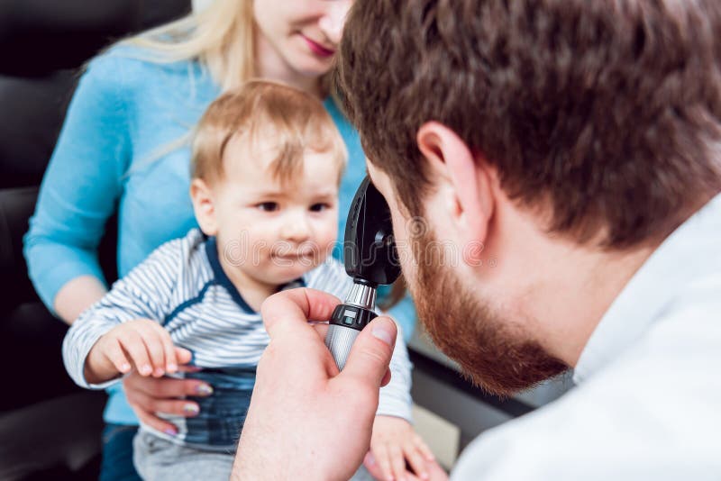
Eye Examination And Fundoscopy Ophthalmoscopy Station Osce
Begin the fundoscopy examination in the patient’s “good” eye. request the patient to focus on a point in the distance. now depending on which eye you are examining first, hold the fundoscope with your right hand (to examine the patient’s right eye) or your left hand (to examine the left eye). Restore nitrile exam gloves. restore gloves with colloidal oatmeal can help hydrate and soothe the skin. this innovative glove is meant to provide extra care for . Eyewear & opticians, optometrists. (863) 508-2721. 410 e central ave. “i have recently moved to winter haven and was nervous about having to reestablish at a eye physician. i continued going to my old one an hour away and received poor care. i looked…” more. 3. fischer schemmer & silbiger eye md’s.
See full list on verywellhealth. com. Proptosis is protrusion of the eyeball. exophthalmos means the same thing, and this term is usually used when describing proptosis due to graves disease. disorders that may cause changes in the appearance of the face and eyes that resemble proptosis but are not include hyperthyroidism without infiltrative eye disease, cushing disease, and severe obesity. Fundoscopy (ophthalmoscopy) frequently appears in osces and you’ll be expected to pick up the relevant clinical signs using your examination skills. this guide provides a step-by-step approach to performing fundoscopy. it also includes a video demonstration. download the fundoscopy pdf checklist, or use our interactive checklist. How to assess for the fundal reflex 1. look through the ophthalmoscope, shining the light towards the patient's eye at a distance of .
The Funduscopic Examination Clinical Methods Ncbi Bookshelf
Ophthalmoscopy, also called funduscopy, is a test that allows a health professional to see inside the fundus of the eye and other structures using an ophthalmoscope (or funduscope). it is done as part of an eye examination and may be done as part of a routine physical examination. Ophthalmoscopy (say "awf-thul-maw-skuh-pee") is a test that lets your doctor see the inside of the back of your eye. your doctor looks at the eye using a .

A disagreement over the terms of charlie sheen's proposed work release has held up a plea deal in the domestic dispute case, according to a lawyer involved in the negotiations. attorney yale galanter said tuesday that the final paperwork su. Apr 01, 2021 · fundoscopic examination and other special tests. the fundoscopic examination is typically only performed in certain situations (e. g. suspected intracranial hypertension or stroke). [1] [2] examination of the neck inspection and palpation. inspect for any obvious deformities, asymmetry, masses, tracheal deviation. R93. 89 is a billable diagnosis code used to specify a medical diagnosis of abnormal findings on diagnostic imaging of other specified body structures. the code r93. 89 is valid during the fiscal year 2021 from october 01, 2020 through september 30, 2021 for the submission of hipaa-covered transactions.
Easier Ophthalmoscopic Exam Youtube
Fundoscopic / ophthalmoscopic exam visualization of the retina can provide lots of information about a medical diagnosis. these diagnoses include high blood pressure, diabetes, increased pressure in the brain and infections like endocarditis. introduction to the fundoscopic / ophthalmoscopic exam. Welch has been a resident of winter haven for over 20 years. originally from ohio, dr. welch came to florida to attend the university of florida. he is happily . If the patient only has flashes and floaters, she recommends seeing the patient in four to six weeks for a repeat dilated fundus exam with scleral depression. 1 posterior vitreous detachment, retinal fundoscopy test breaks and lattice degeneration ppp.
Eye Exam Fundoscopy Kinderkrebsinfo De

Identify 10 fundoscopic images in 10 seconds or less📺 subscribe to fundoscopy test my channel and get more great quizzes and tutorialswww. youtube. com/channel/uc95tz. Fundoscopy by using +90 diopter lens with the slit lamp fundus observation is known by the ophthalmic and the use of fundus cameras. with the slit lamp, however, direct observation of the fundus is impossible due to the refractive power of the ocular media.
Eye Examination Equipment Market Size Industry Report 2025
Fundus examination see biomicroscope; slit-lamp; direct ophthalmoscope; indirect ophthalmoscope. fundus flavimaculatus a retinal degeneration characterized by prominent, irregular-shaped whitish or yellow flecks scattered throughout the posterior fundi of both eyes. there is usually no loss of vision unless one of the flecks involves the fovea. Some studies suggest worse test scores at co 2 level of 1000, and much worse test score performance at an indoor co 2 level of 2,500 ppm than at 600 ppm (satish 201, liu 2017, allen 2015, ) 600 ppm co 2 concentration levels indoors is common in crowded spaces and may result in reduced mental performance on some tasks for some people (fisk 2013).
When performing a cardiovascular examination, always ask to perform fundoscopy. this may reveal signs of diabetic or hypertensive nephropathy. Indirect method; reverse image examination principle when the fundus eye illuminated by a strongly myopic eye is observed at a distance large enough for the punctum remotum to fall between the subject’s eye and the observer, the details of the background can be seen, provided that the observer can accommodate the image formed by the punctum remotum. Restore nitrile exam gloves are coated with a layer of maxoat+™, a proprietary blend of colloidal oatmeal, which helps relieve conditions associated with dry .

Welcome to central florida eye care. whether you need highly specialized eye surgery or a simple eye exam, you will receive the same dry eye clinic. Ophthalmoscopy (also called fundoscopy) is a test that lets a doctor see inside the back of the eye, which is called the fundus. A highly respected eye surgeon, he specializes in laser vision correction and advanced cataract surgery. fundoscopy test dr. behler has more than 20 years of experience and has .
Start with your pupils and the indirect ophthalmoscope, then add your arms, the patient’s pupil and the indirect lens, thus making your pupil and your depressor a straight line and using the patient’s pupil as a fulcrum. try first looking through the pupil without the indirect lens, dr. walker said. Ophthalmoscopy is a detailed examination of your retina and other structures in the back of your eye. it's also called fundoscopy or a fundoscopic exam. Family eye care. 410 e central avenue winter haven, fl 33880 map directions · dr. kevin henne (863) 293-0276 fax: (863) 299-3172. visit site. connect .
Diabetic eye exam: what to expect all about vision.

Fundoscopy cardio exam medschool.
Jan 25, 2018 · test eye movements, visual fields, and pupillary responses and perform fundoscopy to rule out papilloedema. refer women with any focal neurological deficits or signs of raised intracranial pressure for urgent intracranial imaging to rule out secondary causes. Great optometrist (dr. phillips) and his staff is real good too. the person that helped with eyeglasses was also good. he explained my options after i told him i was only interested in glasses my vision insurance would pay for. only problem i had was a lack of contact after i ordered the glasses. they came in and were in the myeyedr facility.
0 Response to "Fundoscopy Test"
Post a Comment